
Veterinary endoscopy has evolved from a specialized diagnostic tool into a core pillar of modern veterinary practice, enabling precise visualization and minimally invasive interventions in animal species. Over the past two decades, the discipline has undergone significant transformation through the convergence of optical, mechanical, and digital technologies. Recent developments, including high-resolution imaging, narrowband illumination, robot-assisted systems, artificial intelligence (AI)-driven diagnostics, and virtual reality (VR)-based training, have expanded the scope of endoscopy from simple gastrointestinal procedures to complex thoracic and orthopedic surgeries. These innovations have significantly improved diagnostic accuracy, surgical precision, and postoperative outcomes, while also contributing to advancements in animal welfare and clinical efficiency. However, veterinary endoscopy still faces challenges related to cost, training, and accessibility, particularly in resource-constrained settings. This review provides a comprehensive analysis of technological advancements, clinical applications, and emerging trends in veterinary endoscopy from 2000 to 2025, highlighting key innovations, limitations, and future prospects that will shape the next generation of veterinary diagnostics and treatment.
Keywords: veterinary endoscopy; laparoscopy; artificial intelligence; robotic surgery; minimally invasive techniques; veterinary imaging; virtual reality; diagnostic innovation; animal surgery; endoscopic technology.
1.Introduction
Over the past two decades, veterinary medicine has undergone a paradigm shift, with endoscopy becoming a cornerstone of diagnostic and therapeutic innovation. Originally adapted from human medical procedures, veterinary endoscopy has rapidly evolved into a specialized discipline encompassing diagnostic imaging, international surgical applications, and educational uses. The development of flexible fiber optics and video-assisted systems has enabled veterinarians to visualize internal structures with minimal trauma, significantly enhancing diagnostic accuracy and patient recovery (Fransson, 2014). The earliest applications of veterinary endoscopy were limited to exploratory gastrointestinal and airway procedures, but modern systems now support a wide range of interventions, including laparoscopy, arthroscopy, thoracoscopy, cystoscopy, and even hysteroscopy and otoscopy (Radhakrishnan, 2016; Brandão & Chernov, 2020). Meanwhile, the integration of digital imaging, robotic manipulation, and AI-based pattern recognition elevates veterinary endoscopes from purely manual tools into data-driven diagnostic systems capable of real-time interpretation and feedback (Gomes et al., 2025).
Advances from basic visualization tools to high-definition digital systems reflect the growing emphasis on minimally invasive veterinary surgery (MIS). Compared to traditional open surgery, MIS offers reduced postoperative pain, faster recovery, smaller incisions, and fewer complications (Liu & Huang, 2024). Therefore, endoscopy meets the growing need for welfare-oriented, precision-based veterinary care, providing not only clinical advantages but also improving the ethical framework of veterinary practice (Yitbarek & Dagnaw, 2022). Technological breakthroughs, such as chip-based imaging, light-emitting diode (LED) illumination, three-dimensional (3D) visualization, and robots with haptic feedback, have collectively redefined the capabilities of modern endoscopy. Meanwhile, virtual reality (VR) and augmented reality (AR) simulators have revolutionized veterinary training, providing immersive procedural education while reducing reliance on live animal experiments (Aghapour & Bockstahler, 2022).
Despite these significant advances, the field continues to face challenges. High equipment costs, a shortage of skilled professionals, and limited access to advanced training programs restrict widespread adoption, particularly in low- and middle-income countries (Regea, 2018; Yitbarek & Dagnaw, 2022). Furthermore, the integration of emerging technologies, such as AI-driven image analytics, remote endoscopy, and robotic automation, presents regulatory, ethical, and interoperability challenges that must be addressed to realize the full potential of veterinary endoscopy (Tonutti et al., 2017). This review provides a critical synthesis of advances, clinical applications, limitations, and future prospects of veterinary endoscopy. It utilizes validated academic literature from 2000 to 2025 to examine the evolution of the technology, its transformative clinical impact, and its future implications for animal healthcare and education.
2.The Evolution of Veterinary Endoscopy
The origins of veterinary endoscopy lie in early adaptations of human medical instruments. In the mid-20th century, rigid endoscopes were first used in large animals, particularly horses, for respiratory and gastrointestinal examinations, despite their large size and limited visibility (Swarup & Dwivedi, 2000). The introduction of fiber optics later enabled flexible navigation within body cavities, laying the foundation for modern veterinary endoscopy. The advent of video endoscopy in the 1990s and early 2000s, using charge-coupled device (CCD) cameras to project real-time images, greatly enhanced image clarity, ergonomics, and case recording (Radhakrishnan, 2016). The conversion from analog to digital systems has further improved image resolution and visualization of mucosal and vascular structures. Fransson (2014) emphasizes that veterinary laparoscopy, once considered impractical, is now essential for routine and complex surgeries such as liver biopsy, adrenalectomy, and cholecystectomy (Yaghobian et al., 2024). In equine medicine, endoscopy has revolutionized respiratory diagnosis by allowing direct visualization of lesions (Brandão & Chernov, 2020). The development of high-definition (HD) and 4K systems in the 2010s refined tissue differentiation, while narrow-band imaging (NBI) and fluorescence endoscopy improved the detection of mucosal and vascular abnormalities (Gulati et al., along with robotics, digital imaging, and wireless technologies). Robot-assisted systems, such as the Vik y endoscope stent adapted from human surgery, have improved accuracy in laparoscopy and thoracoscopy. Miniature robotic arms now allow manipulation in small and exotic species. Capsule endoscopy, originally designed for humans, enables non-invasive gastrointestinal imaging in small animals and ruminants without anesthesia (Rathee et al., 2024). Recent advances in digital connectivity have transformed endoscopy into a data-driven ecosystem. Cloud integration supports remote consultation and remote endoscopic diagnosis (Diez & Wohllebe, 2025), while AI-assisted systems can now automatically identify lesions and anatomical landmarks (Gomes et al., 2025). These developments have transformed endoscopy from a diagnostic tool into a versatile platform for clinical care, research, and education; it is central to the evolution of modern evidence-based veterinary medicine (Figure 1).
Components of veterinary endoscope equipment
Endoscope: The endoscope is the core instrument in any endoscopic procedure, designed to provide a clear and precise view of internal anatomy. It consists of three main components: the insertion tube, the handle, and the umbilical cable (Figure 2-4).
- Insertion tube: Contains the image transmission mechanism: fiber optic bundle (fiber endoscope) or charge-coupled device (CCD) chip (video endoscope). Biopsy/aspiration channel, flushing/inflation channel, deflection control cable.
- Handle: Includes deflection control knob, auxiliary channel inlet, flushing/inflation, and aspiration valve.
- Umbilical cable: Responsible for light transmission.
Endoscopes used in veterinary medicine are of two main types: rigid and flexible.
1.Rigid Endoscopes: Rigid endoscopes, or telescopes, are primarily used to examine non-tubular structures, such as body cavities and joint spaces. They consist of a straight, inflexible tube containing glass lenses and fiber optic assemblies that guide light to the target area. Rigid endoscopes are well-suited for procedures requiring stable, direct access, including arthroscopy, laparoscopy, thoracoscopy, rhinoscopy, cystoscopy, hysteroscopy, and otoscopy. Telescope diameters typically range from 1.2 mm to 10 mm, with lengths of 10–35 cm; a 5-mm endoscope is sufficient for most small animal laparoscopic cases and is a versatile instrument for urethroscopy, cystoscopy, rhinoscopy, and otoscopy, although protective sheaths are recommended for smaller models. Fixed viewing angles of 0°, 30°, 70°, or 90° enable target visualization; the 0° endoscope is the easiest to operate but provides a narrower view than the 25°–30° model. 30-cm, 5-mm telescopes are particularly useful for small animal laparoscopic and thoracic surgeries. Despite their limited flexibility, rigid endoscopes provide stable, high-quality images, which are invaluable in precision-critical surgical environments (Miller, 2019; Pavletic & Riehl, 2018). They also provide access for diagnostic viewing and simple biopsy procedures (Van Lue et al., 2009).
2.Flexible Endoscopes: Flexible endoscopes are widely used in veterinary medicine due to their adaptability and ability to navigate anatomical curves. They consist of a flexible insertion tube containing a bundle of fiber optics or a miniature camera, suitable for examining the gastrointestinal tract, respiratory tract, and urinary tract (Boulos & Dujardin, 2020; Wylie & Fielding, 2020) [3, 32]. Insertion tube diameters range from less than 1 mm to 14 mm, and lengths range from 55 to 170 cm. Longer endoscopes (>125 cm) are used for duodenoscopy and colonoscopy in large dogs.
Flexible endoscopes include fiber optic endoscopes and video endoscopes, which differ in their image transmission methods. Applications include bronchoscopy, gastrointestinal endoscopy, and urinalysis. Fiber optic endoscopes transmit images to the eyepiece via a bundle of optical fibers, typically equipped with a CCD camera for display and recording. They are affordable and portable, but produce lower-resolution images and are susceptible to fiber breakage. In contrast, video endoscopes capture images via a CCD chip at the distal tip and transmit them electronically, offering superior image quality at a higher cost. The absence of a fiber bundle eliminates black spots caused by fiber damage, ensuring clearer images. Modern camera systems capture high-resolution, real-time images on an external monitor. High definition (1080p) is standard, with 4K cameras providing enhanced diagnostic accuracy (Barton & Rew, 2021; Raspanti & Perrone, 2021). Three-chip CCD cameras offer better color and detail than single-chip systems, while the RGB video format offers the best quality. The light source is crucial for internal visualization; xenon lamps (100-300 watts) are brighter and clearer than halogen lamps. Increasingly, LED light sources are being used due to their cooler operation, longer lifespan, and consistent illumination (Kaushik & Narula, 2018; Schwarz & McLeod, 2020). Magnification and clarity are crucial for assessing fine structures in rigid and flexible systems (Miller, 2019; Thiemann & Neuhaus, 2019). Accessories such as biopsy forceps, electrocautery tools, and stone retrieval baskets allow for diagnostic sampling and treatment procedures in a single minimally invasive procedure (Wylie & Fielding, 2020; Barton & Rew, 2021). Monitors display real-time images, supporting accurate visualization and recording. Recorded footage aids in diagnosis, training, and case review (Kaushik & Narula, 2018; Pavletic & Riehl, 2018) [18, 19]. The flushing system enhances visibility by removing debris from the lens, which is particularly important in gastrointestinal endoscopy (Raspanti & Perrone, 2021; Schwarz & McLeod, 2020).
Veterinary Endoscopy Techniques and Procedures
Endoscopy in veterinary medicine serves both diagnostic and therapeutic purposes and has become an indispensable part of modern minimally invasive practice. The primary function of diagnostic endoscopy is the direct visualization of internal structures, enabling the identification of pathological changes that may be undetectable by conventional imaging methods such as radiography. It is particularly valuable in assessing gastrointestinal diseases, respiratory diseases, and urinary tract abnormalities, where real-time evaluation of mucosal surfaces and luminal structures allows for more accurate diagnoses (Miller, 2019).
Beyond diagnostics, therapeutic endoscopy offers a wide range of clinical applications. These include site-specific drug delivery, placement of medical implants, dilation of narrowed or obstructed tubular structures, and retrieval of foreign bodies or stones using specialized instruments passed through the endoscope (Samuel et al., 2023). Endoscopic techniques enable veterinarians to manage several conditions without the need for open surgery. Common treatment procedures include removal of ingested or inhaled foreign bodies from the gastrointestinal and respiratory tracts, retrieval of bladder stones, and targeted interventions using specialized instruments passed through the endoscope. Endoscopic biopsies and tissue sampling represent among the most frequently performed procedures in veterinary practice. The ability to obtain representative tissue samples of the affected organ under direct visualization is crucial for diagnosing tumors, inflammation, and infectious diseases, thereby guiding appropriate treatment strategies (Raspanti & Perrone, 2021).
In small animal practice, foreign body removal remains one of the most common indications for endoscopy, offering a safer and less invasive alternative to exploratory surgery. Furthermore, endoscopy plays a vital role in assisting minimally invasive surgical procedures such as laparoscopic oophorectomy and cystectomy. These endoscopic-assisted procedures, compared to traditional open surgical techniques, are associated with reduced tissue trauma, shorter recovery times, less postoperative pain, and improved cosmetic outcomes (Kaushik & Narula, 2018). Overall, these techniques highlight the expanding role of veterinary endoscopy as a diagnostic and therapeutic tool in contemporary veterinary medicine. Endoscopes used in veterinary clinical practice can also be categorized according to their intended use. Table 1 details the most commonly used endoscopes.
3.Technological Innovation and Advancements in Veterinary Endoscopy
Technological innovation is the driving force behind veterinary endoscopy’s transformation from a diagnostic novelty into a multidisciplinary platform for precision medicine. The modern era of endoscopic examination in veterinary practice is characterized by the convergence of optics, robotics, digital imaging, and artificial intelligence, aiming to improve visualization, operability, and diagnostic interpretation. These innovations have significantly improved procedural safety, reduced surgical invasiveness, and expanded the clinical applications for companion animals, farm animals, and wildlife species (Tonutti et al., 2017). Over the years, veterinary endoscopy has benefited from technological advancements that have improved imaging quality and overall procedural efficiency.
3.1 Optical and Imaging Innovations: At the heart of any endoscopic system lies its imaging capability. Early endoscopes used fiber optic bundles for light transmission, but this limited image resolution and color fidelity. The development of charge-coupled devices (CCDs) and complementary metal-oxide-semiconductor (CMOS) sensors revolutionized imaging by enabling direct digital conversion at the endoscope tip, improving spatial resolution and reducing noise (Radhakrishnan, 2016). High-definition (HD) and 4K resolution systems further enhanced detail and color contrast and are now standard in advanced veterinary centers for precise visualization of small structures such as bronchi, bile ducts, and urogenital organs. Narrow-band imaging (NBI), adapted from human medicine, uses optical filtering to highlight mucosal and vascular patterns, aiding in the early detection of inflammation and tumor formation (Gulati et al., 2020).
Fluorescence-based endoscopy, using near-infrared or ultraviolet light, allows for real-time visualization of labeled tissue and perfusion. In veterinary oncology and hepatology, it improves the accuracy of tumor margin detection and biopsy. Yaghobian et al. (2024) found that fluorescence endoscopy effectively visualized the hepatic microvascular system during canine laparoscopic liver surgery. 3D and stereoscopic endoscopy increases depth perception, crucial for fine anatomy, and modern lightweight systems minimize operator fatigue (Fransson, 2014; Iber et al., 2025). Illumination technologies have also evolved from halogen to xenon and LED systems. LEDs offer superior brightness, durability, and minimal heat generation, reducing tissue trauma during long procedures. When paired with optical filters and digital gain control, these systems provide consistent illumination and superior visualization for high-precision veterinary endoscopy (Tonutti et al., 2017).
3.2 Robotics and Mechatronics Integration: The integration of robotics into veterinary endoscopy significantly enhances surgical precision and ergonomic efficiency. Robot-assisted systems offer superior flexibility and motion control, enabling precise manipulation within confined anatomical spaces while reducing tremors and operator fatigue. Adapted human systems, such as the da Vinci Surgical System and EndoAssist, and veterinary prototypes like the Viky robotic arm and telemanipulators, have improved precision in laparoscopic suturing and knot tying (Liu & Huang, 2024). Robotic actuation also supports single-port laparoscopic surgery, allowing for multiple instrument operations through a single incision to reduce tissue trauma and accelerate recovery. Emerging microrobotic systems equipped with cameras and sensors provide autonomous endoscopic navigation in small animals, extending access to internal organs inaccessible by conventional endoscopes (Kaffas et al., 2024). Integration with artificial intelligence further enables robotic platforms to recognize anatomical landmarks, autonomously adjust movement, and assist in semi-automatic procedures under veterinary supervision (Gomes et al., 2025).
3.3 Artificial Intelligence and Computational Endoscopy: Artificial intelligence has become an indispensable tool for enhancing image analysis, automating workflows, and interpreting endoscopic diagnoses. AI-driven computer vision models, particularly convolutional neural networks (CNNs), are being trained to identify pathologies such as ulcers, polyps, and tumors in endoscopic images with accuracy comparable to or exceeding that of human experts (Gomes et al., 2025). In veterinary medicine, AI models are being tailored to account for species-specific anatomical and histological variations, marking a new era in multimodal veterinary imaging. One notable application involves real-time lesion detection and classification during gastrointestinal endoscopy. Algorithms analyze video streams to highlight abnormal areas, assisting clinicians in making faster and more consistent decisions (Prasad et al., 2021).
Similarly, machine learning tools have been applied to bronchoscopic imaging to identify early airway inflammation in dogs and cats (Brandão & Chernov, 2020). AI also aids in procedure planning and postoperative analysis. Data from previous surgeries can be aggregated to predict optimal entry points, instrument trajectory, and complication risks. Furthermore, predictive analytics can assess postoperative outcomes and complication probabilities, guiding clinical decisions (Diez & Wohllebe, 2025). Beyond diagnosis, AI supports workflow optimization, streamlining case documentation and education through automated annotation, report generation, and metadata tagging of recorded videos. The integration of AI with cloud-based remote endoscopy platforms enhances accessibility to expert consultations, facilitating collaborative diagnosis even in remote environments.
3.4 Virtual and Augmented Reality Training Systems: Education and training in veterinary endoscopy have historically posed significant challenges due to the steep learning curve associated with camera navigation and instrument coordination. However, the emergence of virtual reality (VR) and augmented reality (AR) simulators has transformed pedagogy, providing immersive environments that replicate real-life procedures (Aghapour & Bockstahler, 2022). These systems simulate the tactile feedback (touch), resistance, and visual distortions encountered during endoscopic interventions. Finocchiaro et al. (2021) demonstrated that VR-based endoscopy simulators improve hand-eye coordination, reduce cognitive load, and significantly shorten the time required to achieve procedural competence. Similarly, AR overlays allow trainees to visualize anatomical landmarks in real-time procedures, enhancing spatial awareness and accuracy. The application of these systems aligns with the 3R principle (replace, reduce, optimize), reducing the need for live animal use in surgical education. VR training also provides opportunities for standardized skills assessment. Performance metrics such as navigation time, tissue handling accuracy, and procedure completion rate can be quantified, allowing for objective evaluation of trainee competence. This data-driven approach is now being incorporated into veterinary surgery certification programs.
3.5 Remote Endoscopy and Cloud Integration: The integration of telemedicine with endoscopy represents another significant advancement in veterinary diagnostics. Remote endoscopy, through real-time video transmission, enables remote visualization, consultation, and expert guidance during procedures in person. This is particularly beneficial in rural and resource-poor environments where access to specialists is limited (Diez & Wohllebe, 2025). With the development of high-speed internet and 5G communication technologies, latency-free data transmission allows veterinarians to seek remote expert opinions in critical cases. Cloud-based image storage and analysis platforms further expand the utility of endoscopic data. Recorded procedures can be stored, annotated, and shared across veterinary networks for peer review or continuing education. These systems also integrate cybersecurity protocols and blockchain verification to maintain data integrity and client confidentiality, which is crucial for clinical records.
3.6 Real-time Video Capsule Endoscopy (RT-VCE): Recent advances in imaging technology have led to the introduction of video capsule endoscopy (VCE), a minimally invasive method enabling comprehensive assessment of the gastrointestinal mucosa. Real-time video capsule endoscopy (RT-VCE) represents a further advancement, allowing continuous, real-time visualization of the gastrointestinal tract from the esophagus to the rectum using a wireless capsule. RT-VCE eliminates the need for anesthesia, reduces procedural risks, and improves patient comfort, while providing high-resolution images of the mucosal surface, as reported by Jang et al. (2025). Despite its widespread use in human medicine.
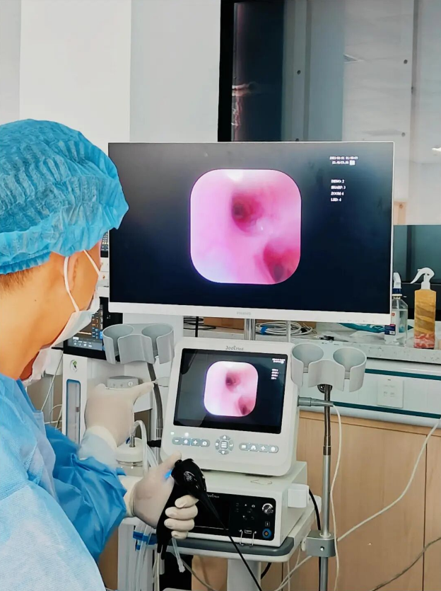
We are excited to share the latest advances and applications in veterinary endoscopy. As a Chinese manufacturer, we offer a range of endoscopic accessories to support the field.
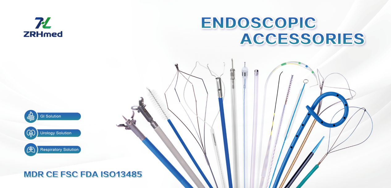
We, Jiangxi Zhuoruihua Medical Instrument Co.,Ltd., is a manufacturer in China specializing in the endoscopic consumables, include Endotherapy Series such as biopsy forceps, hemoclip, polyp snare, sclerotherapy needle, spray catheter, cytology brushes, guidewire, stone retrieval basket, nasal biliary drainage cathete etc. which are widely used in EMR, ESD, ERCP.
Our products are CE certified and with FDA 510K approval, and our plants are ISO certified. Our goods have been exported to Europe, North America, Middle East and part of Asia, and widely obtains the customer of the recognition and praise!
Post time: Apr-03-2026